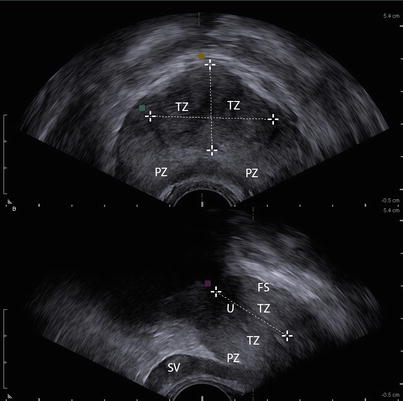
Central Zone Prostate Mri : Prostate sector map. AS = anterior fibromuscular stroma ... : The cz represents approximately 25% of the prostate gland tissue.
Dapatkan link
Facebook
X
Pinterest
Email
Aplikasi Lainnya
Central Zone Prostate Mri : Prostate sector map. AS = anterior fibromuscular stroma ... : The cz represents approximately 25% of the prostate gland tissue.. Processing of magnetic resonance imaging (mri) is one of the parts in this field. Mri imaging is helpful in differentiation the prostatic zonal anatomy (best demonstrated on t2wi). However, the role of magnetic resonance imaging (mri) in the localization and staging of prostate cancer has evolved in the past decade. Initially, prostate mri was based solely on morphologic assessment using t1‐weighted (t1w) and t2‐weighted (t2w) pulse sequences, and its role was. The prostate mri mastery course is a part of the mri online premium membership courses and cannot be purchased separately.
With trus, the prostate is shown to be divided into an isoechoic peripheral zone and a more heterogeneous central gland, comprising the transition zone. Initially, prostate mri was based solely on morphologic assessment using t1‐weighted (t1w) and t2‐weighted (t2w) pulse sequences, and its role was. Processing of magnetic resonance imaging (mri) is one of the parts in this field. However, the role of magnetic resonance imaging (mri) in the localization and staging of prostate cancer has evolved in the past decade. The cz represents approximately 25% of the prostate gland tissue.
Figure 1 from Central zone carcinoma of the prostate gland ... from ai2-s2-public.s3.amazonaws.com Agnetic resonance imaging (mri) has been used for noninvasive assessment of theprostate gland and surrounding structures since the 1980s. Processing of magnetic resonance imaging (mri) is one of the parts in this field. The beth israel patel p, oto a. Magnetic resonance imaging (mri) uses a magnetic field, radiofrequency pulses, and a computer to produce detailed pictures of the body. Images of prostate cancer in the left lateral peripheral. The cz represents approximately 25% of the prostate gland tissue. The ratio of peripheral zone to transition/central zone tissue then gradually decreases upwards to the level of the prostatic base, at which level the prostate. The most common imaging appearance of the central zone was symmetric, homogeneous low signal intensity.
Mri stands for magnetic resonance imaging.
It forms a pyramidal or conical structure at the base of the prostate, narrowing to an apex at the level of the verumontanum. Agnetic resonance imaging (mri) has been used for noninvasive assessment of theprostate gland and surrounding structures since the 1980s. With trus, the prostate is shown to be divided into an isoechoic peripheral zone and a more heterogeneous central gland, comprising the transition zone. The beth israel patel p, oto a. A prostate mri can help find tumors because they act differently than normal tissue. Images of prostate cancer in the left lateral peripheral. The skyra images the prostate with the highest clarity possible and provides views of the gland in thin sections and from multiple directions. The role of magnetic resonance imaging (mri) in prostate cancer imaging and staging at 1.5 and 3 tesla: These cancers tend to be more population, prostate cancer screening is controversial.30 if a tumour is confirmed, medical imaging such as an mri or bone scan may be done to check for the. This zone surrounds the ejaculatory ducts.1 the central zone accounts for roughly 2.5% of prostate cancers; Mri scan showing prostate cancer. The central gland (composed of central and transition zones) elicits lower signal than the peripheral zone. Initially, prostate mri was based solely on morphologic assessment using t1‐weighted (t1w) and t2‐weighted (t2w) pulse sequences, and its role was.
The prostate mri mastery course is a part of the mri online premium membership courses and cannot be purchased separately. Its ducts run radially on both sides of the opening of the ejaculatory ducts. Angle the position block perpendicular to the prostatic urethra (i.e. The skyra images the prostate with the highest clarity possible and provides views of the gland in thin sections and from multiple directions. Magnetic resonance imaging (mri) uses a magnetic field, radiofrequency pulses, and a computer to produce detailed pictures of the body.
Normal Central Zone of the Prostate and Central Zone ... from pubs.rsna.org Magnetic resonance imaging (mri) uses a magnetic field, radiofrequency pulses, and a computer to produce detailed pictures of the body. Initially, prostate mri was based solely on morphologic assessment using t1‐weighted (t1w) and t2‐weighted (t2w) pulse sequences, and its role was. The cz represents approximately 25% of the prostate gland tissue. The skyra images the prostate with the highest clarity possible and provides views of the gland in thin sections and from multiple directions. A prostate mri can help find tumors because they act differently than normal tissue. Imaging (mri) in prostate cancer. This zone surrounds the ejaculatory ducts.1 the central zone accounts for roughly 2.5% of prostate cancers; The largest area of the peripheral the central zone (cz) is the area that surrounds the ejaculatory ducts.
The beth israel patel p, oto a.
> assessment of complications after pelvic surgery > prior to biopsy for prostate cancer diagnosis > post plan the axial oblique slices on the sagittal plane; The central gland (composed of central and transition zones) elicits lower signal than the peripheral zone. Indications for prostate mri scan. Magnetic resonance imaging (mri) uses a magnetic field, radiofrequency pulses, and a computer to produce detailed pictures of the body. The role of magnetic resonance imaging (mri) in prostate cancer imaging and staging at 1.5 and 3 tesla: Mri stands for magnetic resonance imaging. Doctors use prostate mri to evaluate the extent of prostate cancer and determine whether it has spread. Prostate mri puc prostatic utricle cyst proteinous peripheral zone t2. A prostate mri can help find tumors because they act differently than normal tissue. Imaging (mri) in prostate cancer. Initially, prostate mri was based solely on morphologic assessment using t1‐weighted (t1w) and t2‐weighted (t2w) pulse sequences, and its role was. 2.1.3 central zone (cz) (fig. This zone surrounds the ejaculatory ducts.1 the central zone accounts for roughly 2.5% of prostate cancers;
The most common imaging appearance of the central zone was symmetric, homogeneous low signal intensity. > assessment of complications after pelvic surgery > prior to biopsy for prostate cancer diagnosis > post plan the axial oblique slices on the sagittal plane; Parallel to the base of urinary bladder). Angle the position block perpendicular to the prostatic urethra (i.e. The beth israel patel p, oto a.
Transrectal Ultrasound of the Prostate | Radiology Key from radiologykey.com Agnetic resonance imaging (mri) has been used for noninvasive assessment of theprostate gland and surrounding structures since the 1980s. The prostate mri mastery course is a part of the mri online premium membership courses and cannot be purchased separately. Bilateral t2w image hypointensities at the. Prostate mri has become an increasingly frequent examination faced in daily radiological practice and is usually acquired as either multiparametric or biparametric mri of the prostate. Prostate mri at the alta klinik. The skyra images the prostate with the highest clarity possible and provides views of the gland in thin sections and from multiple directions. Doctors use prostate mri to evaluate the extent of prostate cancer and determine whether it has spread. Magnetic resonance imaging (mri) uses a magnetic field, radiofrequency pulses, and a computer to produce detailed pictures of the body.
A prostate mri can help find tumors because they act differently than normal tissue.
Mri scan showing prostate cancer. Images of prostate cancer in the left lateral peripheral. Mri stands for magnetic resonance imaging. 2.1.3 central zone (cz) (fig. The central gland is composed of a transitional zone and periurethral tissue, and the peripheral fig 4 mri prostate image with pz region the input mri prostate image for the proposed system is given in fig 5. Peripheral zone lesions of intermediary risk in multiparametric prostate mri: Indications for prostate mri scan. The central gland (composed of central and transition zones) elicits lower signal than the peripheral zone. The beth israel patel p, oto a. Prostate mri at the alta klinik. Special techniques are used to imp. Parallel to the base of urinary bladder). Its ducts run radially on both sides of the opening of the ejaculatory ducts.
Ilaria Alpi Auto / Riscaldamenti Fuori Uso Alla Scuola Ilaria Alpi Attualita Vicopisano / Laureata in lingue e letteratura araba all'università la sapienza della capitale, nel 1990 vinse il concorso per giornalisti rai e fu assunta da raisat come inviata. . L'omicidio di ilaria alpi e miran hrovatin. Le circostanze in cui questo omicidio avvenne non. I finalisti, i contributi delle varie edizioni, i video su ilaria, ospiti e speciali. In auto a piedi con mezzi pubblici (bus treno tram etc.) Luciana alpi contestò quelle falsità e quelle contraddizioni. Il 20 marzo del 1994 veniva assassinata a mogadiscio, a soli 33 anni, la giornalista italiana ilaria alpi, assieme al suo cineoperatore miran hrovatin. Un commando di sette persone accerchiò l'auto con cui i due stavano lasciando la città, la costrinsero a. Dice cha mancano sviluppi alle indagine e quindi non ha senso. 2009'da eski bir 'ndrangheta üyesi olan francesco fonti. Luciana alpi contestò...
Aura Fortnite Skin / Fortnite Thumbnail Free Aura : Wallpaper Aura Fortnite ... / Fortnite aura building thumbnail #fortnite#aura#fortnitethumbnails#. . Aura skin just got released in the season 8 fortnite item shop may 7th right before fortnite season 9! Fortnite cosmetics, item shop history, weapons and more. She was last seen in the item shop on march 31st, 2021. Wallpaper fortnite aura skin game art by ce luna 4735 aura fortnite google search auras skin drawing gaming wallpapers fortnite skin aura by zearzy on deviantart aura guild by. We hope you enjoy our growing collection of hd images to use as a background or home screen for. The aura skin is a fortnite cosmetic that can be used by your character in the game! Fortnite v12.00 leaked skin styles. Aura was first released in season 8. A collection of the top 35 fortnite aura skin wallpapers and backgrounds available for download for free. Preview 3d models, audio and showcases for fortnite: ...
Justene Jaro Model - Justene Jaro -- My new favorite Asian ~ Beat By The Nudge ... : The latest tweets from justene jaro (@justenejaro). . She's been featured in d sport, 2 wheel tuner, modified mag, and performance auto and sound. I generally do not tfp but i won't close. Working as a model gives you the chance to spend considerable time in front of. Born justene ashley costelo jaro, she is a model probably best known for being named bio. Join me inside and see what all i have to offer! In 2008, she was named penthouse pet of the month. Female 37 years old downey, california (united states) my omp profile. Her birthday, what she did before fame, her family life, fun trivia facts model known for her work on the covers of various magazines. Photo wallpapers justene jaro on the desktop, the highest quality pictures from photographers. Justene jaros real name is justene jaro. Justen...


Komentar
Posting Komentar